Darkfield Microscopy – The River of Life and Your Health
Your bloodstream is your river of life, delivering oxygen, nutrients, and vital healing agents throughout your body. It not only sustains your health but also acts as your body’s natural detoxification system, carrying cellular waste to the liver and kidneys for elimination. But what if this life-giving river became sluggish, murky, or overloaded with toxins?
The truth is, illness begins at the cellular level—often long before symptoms appear. Conventional blood tests may come back “normal” even when underlying health issues are developing. However, Darkfield Live Cell Microscopy provides a real-time, high-definition look at your blood, offering early detection of imbalances, deficiencies, and abnormalities that traditional tests miss.
The Power of Darkfield Microscopy
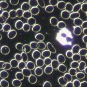
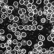
Unlike standard microscopes that use direct light—often washing out crucial details—Darkfield Microscopy uses a unique illumination technique, causing living blood components to glow against a dark background. This cutting-edge technology magnifies your blood 1,500 times and displays it live on a screen, allowing for a deep, dynamic analysis of your health at the cellular level.
By observing your blood in real-time, practitioners can identify:
🔬 Distorted red blood cells – A sign of oxidative stress or nutritional deficiencies.
🛡️ White blood cell vitality – Healthy white blood cells should be active and responsive, not sluggish or damaged. Their condition can reveal how well your immune system is functioning.
❤️ Sticky platelets and clotting risks – Clumped platelets contribute to arterial plaque buildup, increasing the risk of heart attack and stroke.
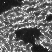
🌬️ Rouleau formation (stacked red blood cells) – When red blood cells clump together, oxygen delivery is compromised, setting the stage for chronic disease, fatigue & brain fog.
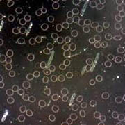
☠️ Toxic buildup and cellular damage – Accumulated toxins, free radicals and upregulated organisms in the blood can damage your cells causing inflammation, fatigue, and a weakened immune system.
A Revolutionary Approach to Health
While conventional lab tests provide static numbers, Darkfield Microscopy offers a real-time, qualitative assessment of your blood, revealing how your body is truly functioning. Experts like Dr. Douglas Brodie, Dr. Katrina Tang, and Dr. Maarten Klatte stress the importance of analyzing the form, movement, and integrity of blood cells, not just measuring them in a lab.
Certified by the renowned Edison Institute of Nutrition, Dr. Kristin specializes in Advanced Live Cell Microscopy, offering deep insights into your health that empower you to take proactive, preventative action. Whether you’re looking to optimize your health, uncover hidden imbalances, or take preventative action, this revolutionary technology can provide the answers you need.
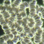
Your Blood Tells a Story—Are You Ready to Listen?
Take control of your health. Darkfield Microscopy provides early detection, unparalleled insight, and the answers you’ve been searching for. Don’t wait for symptoms, see what your blood reveals about your health now. Schedule your analysis today!
